
An ecg components lab notebook is the working record used in teaching and clinical training labs to capture how ECG signals are collected, measured, and interpreted. It connects electrode placement, acquisition settings, waveform identification, and interval measurements into one traceable record that can be reviewed, graded, and reproduced.
In practice, this notebook is where students and supervisors verify correct technique, consistent measurements, and sound interpretation of P waves, QRS complexes, T waves, segments, and intervals. When maintained properly, it supports learning accuracy, assessment fairness, and readiness for real clinical ECG workflows.
What is an ECG lab notebook and what are ECG components?
An ECG lab notebook is a structured record used to document how an electrocardiogram was recorded, measured, and interpreted during a practical session.
-
It captures setup details, raw traces, measurements, and interpretations.
-
It serves as evidence of correct procedure and analytical work.
-
It supports grading, review, and later clinical training reference.
What is an ECG lab notebook in academic and clinical training?
An ECG lab notebook is a formal record of ECG experiments and observations completed by a learner.
-
It is used in physiology, nursing, biomedical, and clinical skills labs.


It documents:
- subject or simulator details
- lead configuration
- acquisition settings
- waveform measurements
It shows how conclusions were reached, not just final answers.
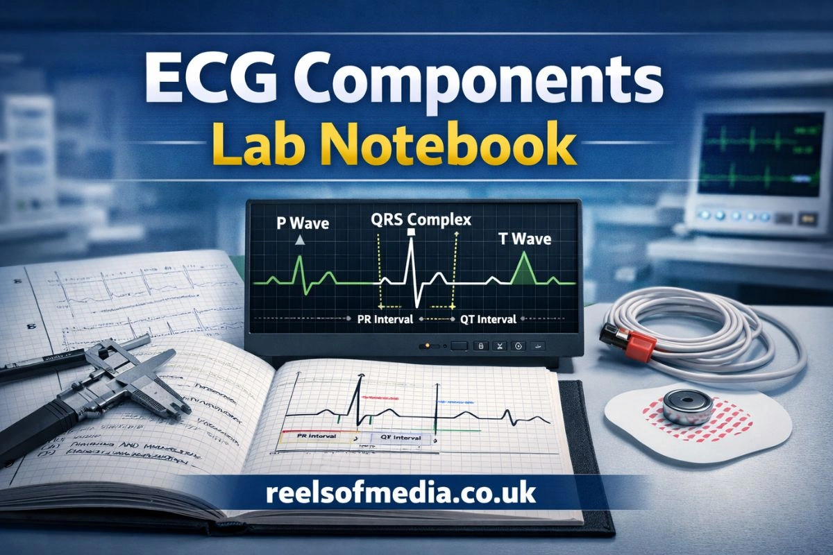
What are the main components of an ECG waveform?
The main components of an ECG waveform are the P wave, QRS complex, and T wave.
-
P wave represents atrial depolarisation.
-
QRS complex represents ventricular depolarisation.
-
T wave represents ventricular repolarisation.
-
These components are the minimum elements expected in most lab records.
What is the difference between ECG waves, segments, and intervals?
Waves are deflections, segments are flat sections, and intervals combine waves and segments.
Waves
- P, Q, R, S, and T deflections
Segments
- PR segment, ST segment
Intervals
- PR interval, QT interval, RR interval
Lab notebooks must clearly separate these three categories.
How does an ECG experiment work in a teaching or training lab?
An ECG experiment follows a standard process of signal acquisition, electrode placement, and digital measurement.
-
The learner prepares the subject or simulator.
-
Electrodes are placed in defined positions.
-
The ECG system records, displays, and stores the waveform.
-
Measurements are taken from the recorded trace.
How is an ECG signal acquired during a lab session?
An ECG signal is acquired by detecting electrical potentials on the skin using surface electrodes.
-
Clean and dry the skin to reduce impedance.
-
Attach electrodes to predefined limb or chest sites.
-
Start recording using the ECG device or software.
-
Confirm signal stability before saving data.
How are leads and electrodes placed for basic ECG recording?
Leads and electrodes are placed using standard limb or chest lead positions.
Typical teaching labs use:
- limb leads (RA, LA, RL, LL)
- selected precordial leads when required
Placement must be:
- symmetrical
- clearly documented
- labelled in the notebook or diagram
How is raw ECG data converted into measurable waveforms?
Raw electrical signals are filtered, amplified, and displayed as time-based waveforms.
The ECG system applies:
- baseline correction
- noise filtering
- signal amplification
The final trace is:
- shown on screen
- exported or printed
- used for manual or software-based measurements
Who is responsible for recording ECG data in a lab notebook?
The primary responsibility for recording ECG data rests with the student performing the experiment.
-
Instructors validate procedures and results.
-
Institutions define minimum documentation standards.
What are the student’s responsibilities during an ECG lab?
Students are responsible for accurate recording, measurement, and basic interpretation.
-
Record setup and electrode placement.
-
Capture representative ECG traces.
-
Measure waves, segments, and intervals.
-
Document any abnormalities or artefacts observed.
What are the instructor or lab supervisor’s responsibilities?
Instructors are responsible for oversight, safety, and data quality control.
-
Verify correct electrode placement.
-
Confirm proper equipment use.
-
Review notebook entries for completeness.
-
Provide feedback on interpretation accuracy.
What documentation is required for assessment or validation?
Assessment documentation includes all information needed to reproduce and verify the experiment.
-
Student identification and date
-
experiment objective
-
equipment and lead configuration
-
recorded ECG trace
-
measurements and brief interpretation
Why is accurate documentation of ECG components important?
Accurate documentation ensures reliable learning, correct interpretation, and fair assessment.
-
It reduces misinterpretation of electrical events.
-
It supports traceability of measurements.
-
It demonstrates professional data handling behaviour.
Why do instructors require ECG components to be clearly labeled?
Clear labels allow instructors to verify that learners correctly identified physiological events.
It confirms understanding of:
- atrial activity
- ventricular activity
- repolarisation phases
It allows rapid checking during grading.
How does accurate recording support learning and interpretation skills?
Accurate recording builds pattern recognition and analytical confidence.
Students learn to:
- distinguish true waves from noise
- measure consistently
- compare results across conditions
It improves readiness for clinical ECG interpretation.
How can poor documentation affect lab results and grading?
Poor documentation directly lowers assessment outcomes and reliability.
Missing labels lead to:
- loss of marks
- unclear interpretations
Inconsistent measurements reduce:
- reproducibility
- credibility of results
What are the core ECG components students must record in a lab notebook?
Students must record all primary waves and the main clinically relevant intervals and segments.
-
P wave
-
QRS complex
-
T wave
-
PR, QT, RR intervals
-
ST and PR segments
How should the P wave be identified and recorded?
The P wave should be identified as the first small deflection before the QRS complex.
- Confirm it precedes ventricular depolarisation.
Measure:
- onset to end duration
- amplitude if required
Note any:
- absence
- abnormal shape
- excessive duration
How should the QRS complex be identified and recorded?
The QRS complex should be identified as the largest rapid deflection group.
Mark:
- Q onset
- R peak
- S end
Measure:
- total QRS duration
Record:
- unusually wide or fragmented complexes
How should the T wave be identified and recorded?
The T wave should be identified as the deflection following the QRS complex.
Mark:
- start and end of the wave
Observe:
- polarity
- symmetry
Note:
- flattened or inverted T waves when present
Which ECG intervals and segments must be measured?
The required intervals and segments include PR, QT, RR intervals and PR and ST segments.
PR interval
- atrial to ventricular conduction time
QT interval
- ventricular depolarisation and repolarisation duration
RR interval
- heart rate calculation
ST and PR segments
- baseline and repolarisation assessment
What are the benefits of a well-prepared ECG components lab notebook?
A well-prepared notebook improves performance, learning continuity, and professional readiness.
-
It creates a reliable reference.
-
It reduces avoidable grading errors.
-
It supports long-term skill development.
How does a structured notebook improve practical exam performance?
A structured notebook provides clear evidence of method and reasoning.
Students can:
- revise real measurements
- recognise common patterns
- avoid repeated procedural mistakes
It supports faster problem solving during assessments.
How does proper documentation help with future clinical training?
Proper documentation mirrors clinical documentation habits.
Learners become familiar with:
- trace interpretation workflows
- standard terminology
- audit-style record keeping
This reduces transition gaps in clinical placements.
How does it support group labs and collaborative experiments?
A well-maintained notebook enables shared understanding and cross-checking.
Team members can:
- verify each other’s measurements
- compare traces
- identify procedural inconsistencies
What are best practices for writing an ECG lab notebook?
Best practice focuses on clarity, consistency, and traceability.
-
Write in real time during the session.
-
Use consistent measurement methods.
-
Avoid rewriting from memory.
How should observations and measurements be formatted?
Observations and measurements should be presented in a structured and readable format.
Use tables for:
- wave durations
- intervals
- amplitudes
Separate:
- raw observations
- interpretation notes
How should units, scales, and time intervals be recorded?
All values must be recorded with correct units and scale references.
Use:
- milliseconds for time
- millivolts for amplitude
Record:
- paper speed or digital time scale
- gain or sensitivity settings
How should annotations and labels be added to ECG traces?
Annotations should be placed directly on the trace or attached as a labelled figure.
Mark:
- P, Q, R, S, T points
- interval boundaries
Keep labels:
- readable
- consistent across figures
What academic or laboratory requirements apply to ECG lab notebooks?
ECG lab notebooks must comply with course, institutional, and data integrity standards.
-
Requirements differ by programme.
-
Core documentation principles remain consistent.
What institutional or course guidelines typically apply?
Most institutions require standardised lab reporting elements.
-
Title and objective
-
method summary
-
recorded data
-
measurements
-
short interpretation
-
student and supervisor identification
What ethical and data integrity rules apply to recorded ECG data?
ECG data must be handled as sensitive physiological information.
-
Do not include identifiable personal data unless authorised.
-
Do not alter traces after recording.
-
Maintain original files or prints for review.
What information must be included for reproducibility?
Reproducibility requires complete procedural and system details.
-
electrode positions
-
lead configuration
-
filter and gain settings
-
recording duration
-
subject or simulator condition
What common mistakes occur when recording ECG components?
Most errors occur during identification, measurement, and documentation.
-
These errors are preventable with structured checks.
How do students misidentify waves and intervals?
Misidentification usually occurs due to baseline noise and poor scaling.
-
Confusing T waves with P waves
-
Selecting incorrect QRS onset
-
Ignoring merged or overlapping deflections
What measurement and calibration errors are most common?
Calibration errors result from incorrect scale assumptions.
-
Using the wrong paper speed
-
Forgetting to check gain settings
-
Measuring on distorted or clipped traces
What documentation and formatting errors reduce marks?
Formatting errors reduce clarity and assessment confidence.
-
Missing labels
-
No units recorded
-
Unclear handwriting or inconsistent tables
What tools and techniques help record ECG components accurately?
Accurate recording relies on standard ECG systems and measurement tools.
-
Digital systems are increasingly used in teaching labs.
-
Manual tools remain important for basic training.
What ECG machines and software are commonly used in teaching labs?
Teaching labs commonly use portable ECG devices and educational software platforms.
-
Desktop or tablet-based ECG acquisition systems
-
Integrated waveform analysis software
-
Simulation ECG platforms for controlled scenarios
What measurement tools are used for interval and amplitude analysis?
Interval and amplitude analysis uses both manual and digital tools.
-
Digital calipers built into ECG software
-
On-screen rulers
-
Printed trace rulers for paper ECGs
How can digital annotations improve ECG lab records?
Digital annotations improve clarity and traceability.
They allow:
- precise marking of wave boundaries
- consistent labelling
- easy revision and review
They reduce transcription errors.
What should be included in an ECG components lab notebook checklist?
A checklist ensures all critical elements are recorded and reviewed.
-
It supports consistency across sessions.
-
It reduces omissions.
What information must be recorded before starting the experiment?
Before recording, setup and context must be documented.
-
experiment objective
-
subject or simulator identifier
-
electrode placement plan
-
lead configuration
-
device and software settings
What must be recorded during ECG signal acquisition?
During acquisition, operational and data details must be captured.
-
time of recording
-
stable ECG traces
-
artefacts or disturbances
-
representative screenshots or printouts
What must be reviewed and submitted after the lab session?
After the session, results and documentation must be finalised.
-
labelled ECG traces
-
completed measurement tables
-
short interpretation notes
-
supervisor verification if required
How do manual and digital ECG lab notebooks compare?
Manual and digital notebooks differ mainly in accuracy, workflow, and review capability.
-
Both can meet academic requirements.
-
Selection depends on programme and infrastructure.
What are the advantages of handwritten ECG lab notebooks?
Handwritten notebooks support foundational skills and manual interpretation.
-
Encourage careful visual inspection
-
Improve understanding of time and scale
-
Simple to implement in basic labs
What are the advantages of digital or electronic lab notebooks?
Electronic notebooks improve accuracy and data management.
-
Automated measurement tools
-
easy file storage and retrieval
-
direct integration of ECG traces
Which approach is more suitable for different learning environments?
The most suitable approach depends on learning objectives.
Manual notebooks suit:
- early training
- basic physiology labs
Digital notebooks suit:
- advanced analysis
- large cohorts
- remote or hybrid teaching
FAQs
What should I include in an ECG components lab notebook?
Include experiment objective, electrode placement, ECG traces, P, QRS, and T wave measurements, intervals and segments, and brief interpretation notes.
How do I accurately identify P, QRS, and T waves in a lab setting?
Look for the first small deflection (P wave), the largest rapid deflection group (QRS complex), and the wave following QRS (T wave). Use consistent scales and labels.
What is the proper method for measuring ECG intervals and segments?
Measure PR, QT, and RR intervals using calibrated time scales, mark segment start and end points, and record values in milliseconds with clear labels.
Why is it important to label ECG components clearly in a lab notebook?
Clear labeling ensures instructors can verify correct identification, supports reproducible results, and reduces grading errors.
How can an ECG components lab notebook improve my practical skills and exam performance?
Maintaining a structured notebook helps you track measurements, identify waveform patterns, and practice consistent interpretation, boosting exam readiness.